Traditional vs. Digital Dental Model Creation: A Practical Pros and Cons Comparison
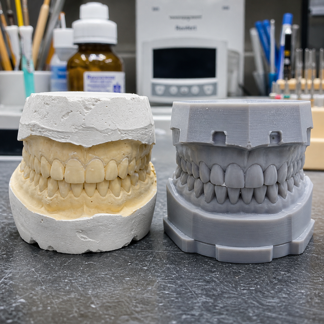
Dental model creation sits at the foundation of nearly every restorative and orthodontic workflow. Whether a technician is fabricating a crown, designing a clear aligner, or planning implant placement, the quality of the model directly shapes the quality of the outcome. For decades, stone and plaster casts made from alginate or polyvinylsiloxane impressions were the only option. Today, intraoral scanners, CAD/CAM software, and 3D-printed dental models have created a genuine fork in the road — and the right path depends heavily on your specific clinical or laboratory context.
What Is Traditional Dental Model Creation?
Traditional dental model creation uses physical impression materials — most commonly alginate or polyvinylsiloxane (PVS) — to capture the patient's dentition, then pours stone or plaster into the negative to produce a solid cast. This analog process has been refined over roughly a century and remains the baseline against which all digital alternatives are measured.
The workflow typically runs as follows: the clinician seats a tray loaded with impression material, waits for it to set (anywhere from 45 seconds to several minutes depending on the material), removes it carefully, and ships or hand-delivers it to the dental laboratory. Lab technicians then mix dental stone, vibrate out air bubbles, and wait for the cast to cure before trimming and articulating it.
Stone casts are dimensionally stable once set, require no software, and can be handled and inspected without a screen. For many practices — particularly those with established lab relationships and high patient volumes — this workflow is deeply familiar and operationally predictable. The consumable costs are low: a bag of alginate runs a few dollars, and dental stone is similarly affordable.
The trade-offs are real, though. Material distortion is a persistent risk — alginate can shrink if not poured promptly, PVS impressions can tear on undercuts, and shipping introduces its own hazards. Physical storage of plaster models consumes significant space over time, and there is no built-in redundancy if a cast is damaged.
What Is Digital Dental Model Creation?
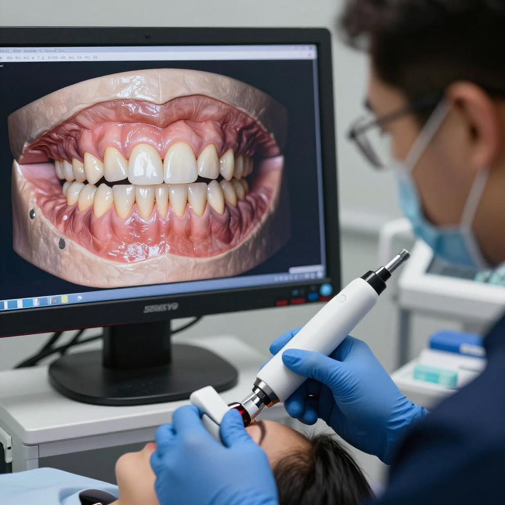
Digital dental model creation replaces physical impression materials with an intraoral scanner that captures a precise 3D point cloud of the dentition, which is then processed into a digital model file — typically an STL or OBJ — ready for CAD/CAM design or direct 3D printing.
The core components of a digital workflow are three: the intraoral scanner (devices like the Medit i700, 3Shape TRIOS, or iTero Element), the CAD software used to design restorations or appliances, and the output method — either milling from a block or, increasingly, resin-based 3D printing. For dental labs and practices focused on model fabrication specifically, 3D printing has become the dominant output path because it handles complex geometries and full-arch cases that milling cannot easily replicate.
Once the scan is complete, the digital file can be sent to a lab within minutes via encrypted cloud transfer, eliminating shipping delays entirely. The lab receives a file they can open, inspect, and print on demand — or archive indefinitely without physical storage requirements.
Accuracy and Clinical Reliability
Both methods can achieve clinically acceptable accuracy, but they fail in different ways and at different points in the workflow. Understanding where each method introduces error is more useful than a blanket claim of superiority for either approach.
Traditional PVS impressions, when taken correctly with proper technique and poured within the recommended time window, can achieve dimensional accuracy within 50–100 microns. The problem is that the error chain is long: impression distortion, pour timing, stone expansion during setting, and trimming errors all compound. A single misstep — say, a torn margin or a delayed pour — can invalidate the model entirely.
Intraoral scanners have improved dramatically since their first clinical iterations. Current-generation devices achieve accuracy figures of 10–30 microns for single-tooth restorations in controlled conditions. Full-arch scanning accuracy, however, remains a more nuanced topic. Scan stitching across a full arch can introduce cumulative error, particularly in the posterior regions, and this has been documented in peer-reviewed literature comparing digital and conventional impressions for complete-arch implant cases.
For a single-tooth crown or a three-unit bridge, digital scanning is arguably more reliable than a conventional impression because it eliminates several error sources simultaneously. For a full-arch implant case requiring passive fit across six or more implants, some clinicians still prefer a conventional open-tray impression or a hybrid approach that verifies the digital scan with a physical check record.
Workflow Efficiency and Turnaround Time
Digital workflows are faster at the communication stage but require more upfront investment in process design. Traditional workflows are slower overall but require almost no infrastructure to operate.
A conventional impression workflow from chairside to model delivery typically spans 24–72 hours when factoring in lab shipping, stone pouring, and curing time. Rush cases can compress this to same-day if the lab is local, but that is the exception. Digital scanning eliminates the physical transport leg entirely — a scan taken at 9 a.m. can be in a lab technician's software queue by 9:05 a.m.
Chairside, a skilled clinician can take a full-arch PVS impression in 5–8 minutes. A full-arch intraoral scan on an experienced operator takes roughly the same time, though the learning curve means early adopters often spend 15–20 minutes per arch until technique stabilizes. Once proficiency is established, scan retake rates tend to drop below those of traditional impressions, which require a complete restart if a margin is missed.
For orthodontic appliance fabrication — aligners, retainers, study models — the digital path offers clear throughput advantages. A lab printing 3D dental models overnight can produce a week's worth of cases in a single batch run, something physically impossible with stone casting.
Cost Considerations — Upfront vs. Long-Term
The cost comparison between traditional and digital model creation is not simply "digital costs more." The more accurate framing is that digital shifts costs from recurring consumables toward upfront capital and maintenance.
A quality intraoral scanner carries a purchase price of $15,000–$35,000 USD, with annual subscription fees for software updates ranging from $1,500–$5,000 depending on the vendor. A dental 3D printer capable of producing accurate models adds another $3,000–$15,000 depending on resolution and build volume, plus resin costs of roughly $80–$150 per liter. These are not trivial numbers for a single-operatory practice.
Traditional impression materials and stone, by contrast, cost a fraction per case — often under $5 in materials for a full-arch impression and pour. But this calculation ignores labor time, storage costs, and the cost of remakes. Impression remakes in a busy practice can run 5–10% of cases; each remake costs chairside time, patient goodwill, and lab fees.
For a high-volume practice or lab producing 30+ models per week, the break-even on digital equipment typically falls within 18–36 months when accounting for reduced remakes, eliminated shipping fees, and faster case throughput. For a low-volume practice doing occasional models, traditional methods may remain more cost-effective indefinitely.
Patient Experience and Comfort
Patients consistently prefer intraoral scanning over traditional impressions, and this preference has measurable effects on practice reputation and case acceptance. That said, the difference matters more for some patient populations than others.
The primary complaint with conventional impressions is the gag reflex — particularly with upper arch alginate trays. Patients with a strong gag response, dental anxiety, or a small mouth can find the process genuinely distressing. Some clinicians use topical anesthetics or distraction techniques, but these add time and don't fully resolve the issue.
Intraoral scanning involves a small wand that moves around the mouth for 3–5 minutes. Most patients find it significantly more comfortable, and many report that seeing their teeth rendered in real time on a screen is genuinely engaging. This visual element also creates a natural opportunity for patient education — pointing out wear patterns, recession, or bite issues on the digital model in ways that a plaster cast sitting on a counter simply cannot replicate.
For pediatric patients or those with special needs, the shorter and less invasive nature of scanning can meaningfully reduce appointment stress. For patients who are already comfortable with traditional impressions and have no gag issues, the experiential difference is less dramatic.
Which Method Is Right for Your Practice or Lab?
The right choice depends on your case mix, volume, budget, and how central model fabrication is to your revenue model. There is no universally correct answer.
Consider traditional impression workflows if your practice does fewer than 10–15 models per month, your lab relationships are well-established and local, or your patient population is largely comfortable with conventional impressions. The capital cost of going digital may not justify the operational savings at low volumes, and the learning curve for intraoral scanning is real — expect 3–6 months before scan quality and speed match your current impression workflow.
Digital workflows — particularly those centered on intraoral scanning and 3D-printed dental models — make strong sense for orthodontic-heavy practices, high-volume restorative labs, or any setting where same-day dentistry or remote lab communication is a priority. The ability to archive every case digitally, with zero physical storage overhead, is also a meaningful operational advantage over time.
Many clinicians land on a hybrid approach: using intraoral scanning for routine restorative and orthodontic cases while retaining the option for conventional PVS impressions in complex implant cases or when scanner access is unavailable. This pragmatic middle ground is increasingly common and reflects the reality that both methods still have a place in a well-equipped practice.
The shift from analog to digital in dental model creation is not a binary switch — it's a spectrum. Where you sit on that spectrum should be driven by your specific clinical needs, not by technology enthusiasm or inertia.
Frequently Asked Questions
Can digital and traditional methods be used together in a hybrid workflow?
Yes, and many practices do exactly this. A common approach is using intraoral scanners for single-unit and orthodontic cases while reserving conventional PVS impressions for full-arch implant cases where scan stitching accuracy is a concern. The two methods are not mutually exclusive, and maintaining both capabilities gives clinicians flexibility across different case types.
How accurate are 3D-printed dental models compared to stone casts?
Current resin-based 3D printers using DLP or MSLA technology can produce dental models accurate to within 50–100 microns — comparable to well-poured stone casts. The key variable is print resolution, resin quality, and post-processing consistency. High-end dental printers from manufacturers like Formlabs, SprintRay, or Asiga routinely meet the accuracy thresholds required for orthodontic appliance fabrication and indirect bonding trays.
What is the learning curve for adopting intraoral scanners?
Most clinicians reach basic proficiency within 4–8 weeks of regular use, but achieving scan quality that consistently matches or exceeds conventional impressions typically takes 3–6 months. The posterior lingual regions and tight contacts are the most common challenge areas. Vendor training programs and in-office coaching from a digital dentistry consultant can significantly compress the learning curve.
Is digital model creation suitable for full-arch cases?
For most full-arch restorative and orthodontic cases, yes. For full-arch implant cases requiring passive fit across multiple implants, the evidence is more mixed. Some studies show acceptable accuracy with current scanner technology; others document cumulative stitching error that affects fit. Many implant specialists use a digital scan for initial planning and a conventional impression or scan body verification for final restoration fabrication.
How are digital dental models stored and shared with labs?
Digital models are stored as STL or PLY files and can be archived on local servers, practice management systems, or cloud-based platforms. Most intraoral scanner ecosystems include integrated cloud portals — 3Shape Communicate, Medit Link, and the iTero portal are widely used examples — that allow encrypted file transfer to any connected laboratory within seconds. Digital archiving eliminates the physical storage problem entirely and provides built-in redundancy that no plaster model can match.