The Role of AI in Enhancing Digital Dental Model Precision
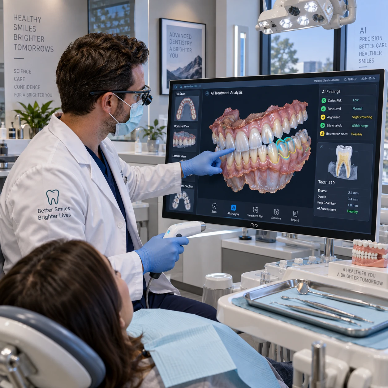
Artificial intelligence is changing how dental teams turn scans into usable digital models. By helping identify tooth surfaces, separate structures, and flag potential data problems, AI can make parts of the modeling process more consistent. Its value, however, depends on the quality of the scan and the judgment of the dental professionals who review the result.
What Digital Dental Models Are—and Why Precision Matters
Digital dental models are three-dimensional representations of a patient’s teeth and, in some workflows, surrounding oral structures. Their precision matters because dentists and dental laboratories use them to assess anatomy, plan treatment, and design restorations or appliances.
Many models begin with data captured by intraoral scanners, which record the surfaces of teeth as the clinician moves the scanner around the mouth. Software combines that data into a 3D dental scan that can be viewed, measured, and used in computer-aided design (CAD). A model may also be created from a scan of a physical impression or cast.
Small defects can have practical consequences. Missing surface data or a distorted tooth margin may make it harder to design a crown that fits as intended. In orthodontic planning, incomplete anatomy can affect measurements or the interpretation of tooth positions. An error in an occlusal surface may also complicate occlusion analysis, which considers how the upper and lower teeth meet.
Precision is not simply a matter of having a smooth-looking model. A clinically useful model must represent relevant anatomy well enough for its intended task. The level of detail needed for a broad treatment discussion may differ from what is required to design a restoration at a specific margin.
How AI Works With Dental Scans
AI works with dental scans by analyzing image or surface data to recognize patterns, identify structures, and assist with preparing a digital model. It can speed up parts of the workflow, but the scan itself remains the source of the information.
An AI system may be trained to distinguish tooth surfaces from soft tissue, detect likely tooth boundaries, or identify areas where scan data appear incomplete. Depending on the software, it can assist with image segmentation, which separates a scan into meaningful regions, and may suggest landmarks or model edits for the operator to review.
The typical process looks like this:
- Capture: An intraoral scanner records a series of images or surface measurements as the clinician scans the mouth.
- Reconstruction: Software aligns overlapping data to build a 3D dental scan. AI may help interpret the data or identify areas that need attention.
- Segmentation and preparation: The system can help label teeth and other structures, remove selected scan artifacts, or prepare a model for CAD.
- Review: A clinician or dental laboratory technician checks whether the result is complete and suitable for its intended use.
AI tools vary. Some make suggestions within a scanning or design application; others automate selected steps. Users should check what the specific product does, what data it needs, and whether its output can be edited. Automation can reduce repetitive work, but it may also make an error less obvious if no one inspects the result.
Where AI Can Improve Model Precision
AI can support digital dental model precision by helping teams interpret scan data, segment anatomy, assess occlusion, and apply repeatable processing rules. These capabilities can make output more consistent, though they cannot restore anatomical details that the scanner never captured.
Scan interpretation and segmentation
AI can help identify tooth boundaries and distinguish teeth from nearby soft tissue. Better segmentation can make it easier to isolate individual teeth for measurement or CAD work. It may also help draw attention to gaps, overlaps, or irregular areas in a scan. The operator still needs to confirm that the software has assigned the correct boundaries, especially around crowded teeth, restorations, or gingival margins.
Occlusion and consistency checks
When upper and lower scans are aligned, software can help analyze how the arches relate and where contacts may occur. AI-assisted occlusion analysis can support review by highlighting potential interferences or areas to inspect. These outputs are aids, not a substitute for clinical assessment of the patient’s bite.
AI may also apply the same processing steps across many cases, reducing variation caused by different users handling routine edits in different ways. That consistency can be useful in a busy clinic or dental laboratory. However, a standardized process can repeat the same mistake if its assumptions do not fit a particular scan. Consistency and correctness are related, but they are not the same.
For a practical review, consider three questions: Is the anatomy captured? Are the boundaries plausible? Does the model suit the task? This quick check helps focus review on the parts most likely to affect downstream work.
Benefits Across Dental Workflows
AI-assisted digital models can support diagnosis, treatment planning, communication, and dental laboratory design by making scan data easier to interpret and prepare. The benefit is strongest when a tool fits an existing workflow and its output is checked before decisions are made.
For diagnosis, clearer segmentation and measurements may help a dentist compare tooth positions or examine a region of interest. For treatment planning, a well-prepared 3D model can provide a shared reference when discussing restorative, orthodontic, or other dental procedures. It helps clinicians inspect anatomy from different angles and explain a proposed plan to a patient.
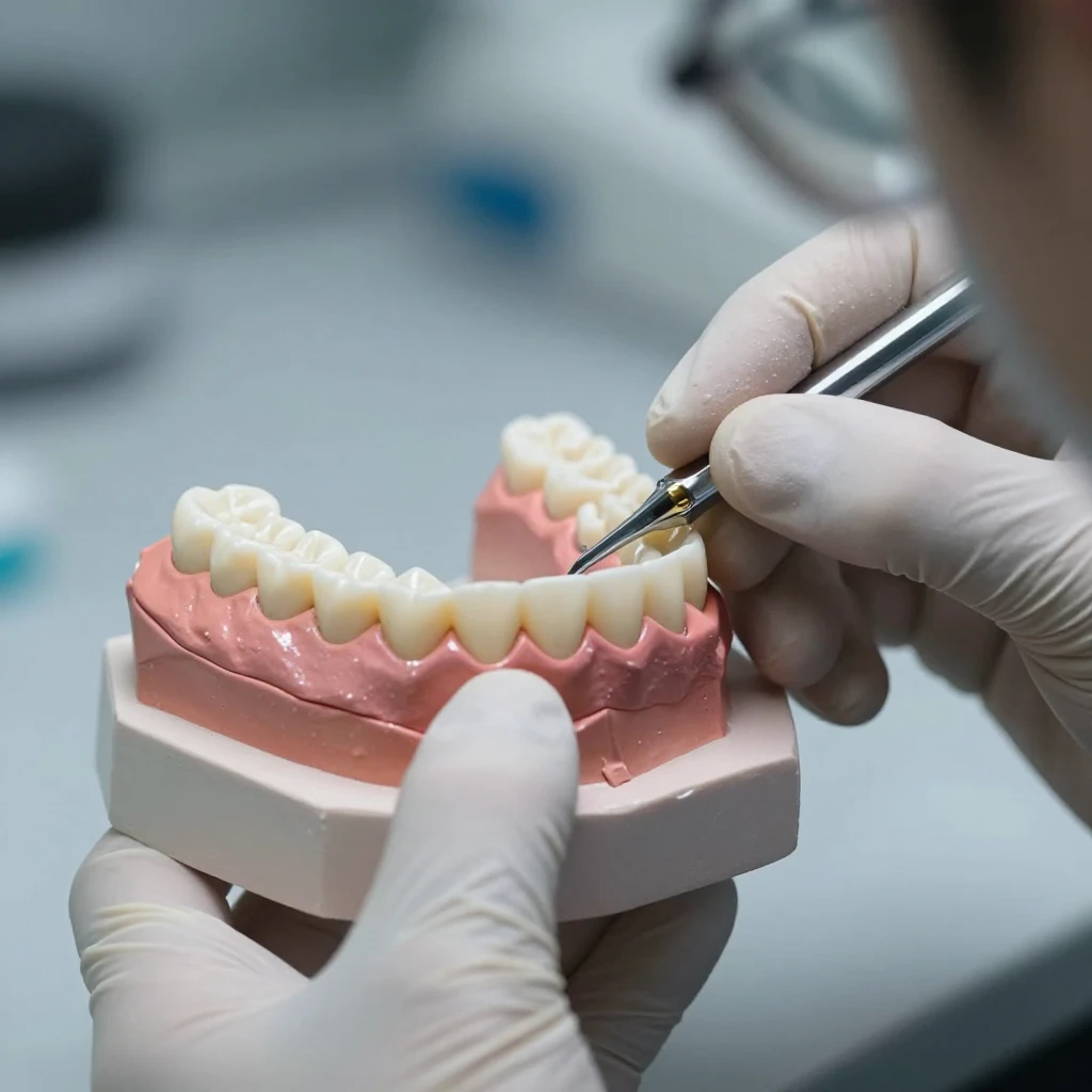
Dental laboratories can use digital models as inputs to CAD software for designing restorations, appliances, and other devices. AI-supported preparation may reduce some repetitive steps, such as separating teeth or identifying areas for review. That can make file handoffs more orderly, but lab technicians still need to confirm margins, contacts, material requirements, and the design’s fit with the prescription.
Digital communication is another practical advantage. A dentist can send a scan and relevant notes to a lab without shipping a physical impression, while both teams can refer to the same file. That convenience depends on clear case information and compatible systems. A clean-looking model without details about the intended restoration or clinical constraints can still lead to avoidable revisions.
Limitations and the Need for Clinical Oversight
AI-generated or AI-assisted models have limitations: poor scan data, complex anatomy, and software assumptions can all affect the result. Clinical validation by a qualified dental professional remains essential before a model informs diagnosis, treatment, or fabrication.
AI cannot reliably infer every surface hidden by saliva, blood, soft tissue, or an obstructed scanner view. If an intraoral scan contains stitching errors, motion artifacts, or missing data, an algorithm may smooth or interpret the defect rather than identify the true anatomy. In complex cases, including crowded teeth or unusual restorations, automated segmentation may need substantial correction.
Data handling also deserves attention. Dental scans are sensitive health information, so practices should understand how a vendor stores, processes, and protects files, and whether data may be used to train or improve its systems. Follow applicable privacy requirements and the organization’s policies rather than assuming that cloud-based processing is automatically appropriate.
Common mistakes to avoid include:
- Accepting a polished model at face value: Smooth surfaces can hide missing or misread anatomy. Inspect critical margins and contact areas before design or clinical use.
- Using an unsuitable scan: AI cannot compensate for incomplete capture. Rescan areas with gaps or distortion when they matter to the case.
- Treating an AI flag as a diagnosis: Alerts and measurements need professional interpretation in the context of the patient and treatment plan.
The central trade-off is straightforward: more automation can reduce routine manual work, but it can also shift effort toward checking exceptions and correcting systematic errors. Teams should preserve a clear review step, particularly for irreversible treatment decisions or devices that must fit precisely.
Integrating AI Into Digital Dentistry
To integrate AI into digital dentistry, start with a specific workflow problem, test the tool on representative cases, and define who reviews its output. A focused evaluation is more useful than adopting software because it promises automation in general.
Before choosing a system, map the current path from scan capture to final model or CAD design. Note where staff spend time, where errors or rework occur, and which checks are already in place. Then evaluate whether the AI feature addresses a real bottleneck, such as segmentation or scan-quality review, without introducing extra file conversions or unclear responsibilities.
- Test varied cases: Include routine scans as well as cases with restorations, crowding, soft-tissue interference, or incomplete capture.
- Compare outputs with reviewed models: Have experienced clinicians or technicians inspect model boundaries and clinically important surfaces.
- Check workflow fit: Confirm compatibility with intraoral scanners, CAD systems, laboratory file formats, and existing review steps.
- Set review rules: Specify which cases require edits, rescan requests, or additional clinical review.
- Reassess over time: Track practical indicators such as correction frequency, remakes, and time spent reviewing, rather than relying only on a vendor’s headline claims.
AI is a good fit when it makes a defined task easier to perform and its limitations are visible to the people using it. It is a poor fit when teams cannot explain how the software reached an output, cannot review that output, or expect the tool to repair inadequate source data.
Frequently Asked Questions
How does AI improve digital dental model accuracy?
AI can help identify anatomy, segment teeth, flag possible scan problems, and standardize selected processing steps. These functions may improve consistency and help users find areas that need review. Accuracy still depends on the original scan and professional validation.
Can AI replace manual review of dental scans?
No. AI can assist with scan interpretation and model preparation, but it can misclassify anatomy or overlook defects. A dentist or qualified dental laboratory professional should confirm that the model is complete and appropriate for its intended use.
What scan data does AI use to create digital models?
AI generally works with data produced by an intraoral scanner or with digitized impressions and casts. Depending on the system, the input may include images, measured surface points, or an assembled 3D dental scan. The software’s documentation explains which formats and data types it accepts.
How can inaccurate scans affect AI-generated models?
Missing, distorted, or poorly aligned scan data can produce an incomplete or misleading digital model. AI may flag some problems, but it cannot reliably reconstruct anatomy that was not captured. Rescanning the affected area is often the appropriate response when the missing detail matters clinically or to lab design.