How Digital Impressions Are Revolutionizing Patient Comfort and Accuracy in Modern Dentistry
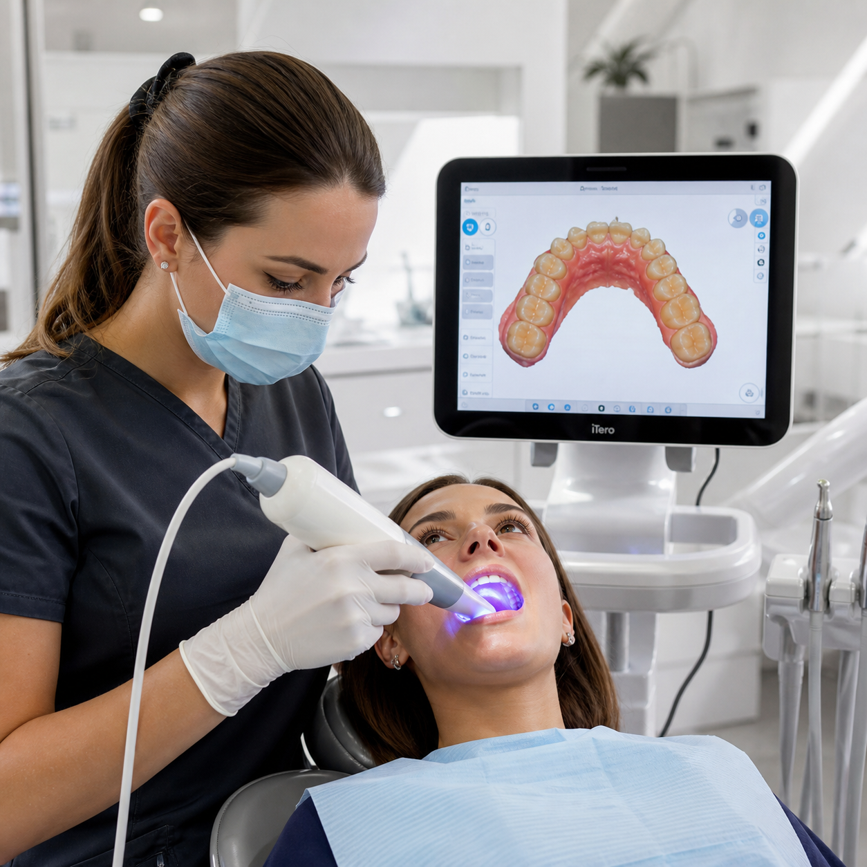
Dental visits have always carried a certain reputation — and not always a flattering one. But one quiet technological shift is changing the experience from the inside out. Digital dental impressions, captured through handheld intraoral scanners, are replacing the cold, uncomfortable trays of putty that patients have tolerated for decades. The result is faster appointments, more precise restorations, and a direct pipeline into 3D printing workflows that produce better-fitting dental models than ever before.
What Are Digital Impressions and How Do They Work?
A digital dental impression is a three-dimensional scan of a patient's teeth and soft tissue, captured using a small handheld device called an intraoral scanner (IOS). Instead of pressing a tray filled with impression material into the mouth, the clinician moves the scanner wand slowly across the teeth, and the device stitches together thousands of images per second into a precise digital model.
The process takes roughly two to five minutes per arch. Once complete, the scan data — typically exported as an STL or OBJ file — transfers directly into CAD/CAM (Computer-Aided Design / Computer-Aided Manufacturing) software. From there, a dental technician or an automated system can design a crown, aligner, or bridge digitally, then send the file to a milling machine or a 3D printer to fabricate the physical restoration or model.
There are no mixing bowls, no setting times, and no physical material to store or ship. The entire capture-to-model process lives in the digital domain until a physical output is needed.
The Problem with Traditional Impressions
Conventional alginate and polyvinyl siloxane (PVS) putty impressions have served dentistry well for over a century — but they come with real limitations that digital alternatives expose clearly.
The most immediate issue is physical discomfort. Traditional impression trays are loaded with viscous material and seated firmly against the teeth, often extending toward the back of the mouth. For patients with a sensitive gag reflex, this can make a routine appointment feel genuinely distressing. Some patients avoid necessary dental work specifically because of this experience.
Beyond comfort, there's the accuracy problem. Impression materials can distort during removal, especially around undercuts or when the patient moves unexpectedly. The physical model then has to be poured in stone, shipped to a lab, and scanned or milled — each step introducing a small margin of error. By the time a crown is fabricated, the cumulative distortion can require a remake, costing both the patient and the practice time and money.
Workflow inefficiency compounds everything. Traditional impressions typically add days to the treatment timeline while physical models travel to and from dental laboratories.
Patient Comfort: A Measurable Difference
Eliminating physical impression material produces a concrete, immediate improvement in the patient experience. Patients no longer have to hold still while a tray sets — a process that can take two to five minutes and feels much longer when you're trying not to gag.
Research published in clinical dental journals consistently shows that patients rate intraoral scanning significantly more comfortable than conventional impressions. One frequently cited finding is that patients with a pronounced gag reflex report near-complete relief when the physical tray is removed from the equation. For pediatric patients and individuals with dental anxiety, this difference can determine whether they return for follow-up care at all.
Chair time also drops. A full-arch digital scan typically takes two to four minutes, compared to the ten to fifteen minutes a traditional impression workflow can occupy when you account for material mixing, setting, removal, and rinsing. Shorter appointments reduce fatigue and anxiety — both of which affect how patients perceive their overall dental experience.
There's also a psychological dimension worth noting: patients can see their own teeth rendered in real time on a chairside screen. That visual transparency builds trust and opens natural conversations about treatment options.
Accuracy and Clinical Precision: What the Technology Delivers
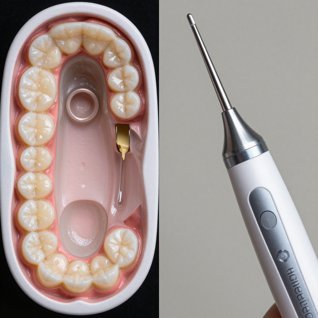
Digital impressions reduce the error chain that exists in every traditional impression workflow. The scan captures tooth geometry directly, without the material distortion, model pouring variables, or shipping damage that can affect physical impressions.
Modern intraoral scanners achieve dimensional accuracy in the range of 10 to 50 microns across a full arch — a level of precision that translates directly into better-fitting restorations. Crowns and bridges fabricated from digital scans require fewer adjustments at delivery, and remake rates drop measurably in practices that have switched from conventional to digital workflows.
The consistency is also worth emphasizing. A physical impression depends on the clinician's technique, the material's mixing ratio, the ambient temperature, and the patient's cooperation. A digital scan standardizes most of those variables. The scanner captures what's there; the software processes it the same way every time.
This doesn't mean digital impressions are error-free. Saliva, blood, and inadequate retraction can still compromise scan quality. But the failure mode is visible immediately — the software flags incomplete or low-confidence regions, and the clinician can rescan that area on the spot rather than discovering the problem days later when a lab calls about a poor-quality impression.
From Scan to 3D-Printed Model: The Digital Workflow
The real power of digital impressions emerges when the scan data connects to a downstream fabrication workflow. Once a scan is complete, the STL file can be sent directly to a CAD/CAM system, where a technician designs the restoration virtually, or to a 3D printer that produces a physical dental model without any lab intermediary.
This is where point-of-care 3D printing becomes significant. Dental practices equipped with in-office printers can produce a physical model, a surgical guide, or a temporary crown within hours of the scan — sometimes within the same appointment. The traditional model, which required sending physical impressions to an external lab and waiting days or weeks for results, is giving way to same-day or next-day delivery.
The digital workflow also enables parallel processing. While the patient is still in the chair, the scan file can be reviewed remotely by a specialist, sent to multiple labs for quoting, or archived permanently in the patient record. Physical models, by contrast, can be lost, broken, or degraded over time.
3D-printed dental models produced from digital impression data are dimensionally consistent with the original scan, which means the restoration designed on that model fits the patient's actual anatomy — not a slightly distorted physical replica of it. That chain of fidelity, from scan to software to printed model to finished restoration, is what makes the digital workflow genuinely superior to its predecessor.
Which Treatments Benefit Most from Digital Impressions?
Digital impressions add value across a wide range of dental treatments, though some applications benefit more than others.
- Crowns and bridges: The most common application. Digital scans eliminate the most error-prone steps in crown fabrication, and same-day crowns milled from ceramic blocks are now routine in digitally equipped practices.
- Orthodontic aligners: Clear aligner systems depend entirely on accurate digital models. Most major aligner manufacturers now require or strongly prefer digital submissions over physical impressions.
- Implant planning: Digital scans integrate with CBCT (cone beam CT) data to produce surgical guides that position implants with sub-millimeter precision.
- Nightguards and retainers: Simple appliances that previously required lab turnaround can now be printed in-office within a day.
- Full-arch restorations: Modern scanners handle full-arch cases reliably, though technique and patient cooperation still matter more at this scale.
Treatments involving deep subgingival margins or complex posterior anatomy can still present challenges for intraoral scanning, and some clinicians prefer supplementing with traditional methods in those specific situations. The technology is strong across most of the clinical range, but it's not a universal replacement for every scenario yet.
What to Expect as Digital Impressions Become the Standard
Digital impressions are already the default in many progressive dental practices, and adoption is accelerating. Industry data from dental technology surveys suggests that intraoral scanner adoption among general dentists has roughly doubled over the past five years, driven by falling hardware costs and the growing expectation from patients — particularly younger ones — that their dental experience should feel modern.
Point-of-care 3D printing is the next frontier. As desktop dental printers become faster, more affordable, and easier to validate for clinical use, the gap between scan and finished product will continue to shrink. Practices that invest in the full digital workflow — scanner, software, and printer — are positioning themselves to offer same-visit restorations as a standard service rather than a premium offering.
For patients, the practical implication is fewer appointments, less discomfort, and restorations that fit better from the start. For dental professionals, it means a more controlled, efficient workflow with lower remake rates and stronger patient retention.
The shift isn't hypothetical. It's already happening in practices around the world, and the gap between digitally equipped clinics and those still using putty trays is becoming harder to ignore.
Frequently Asked Questions
Are digital impressions covered by dental insurance?
Coverage depends on the insurer and the specific plan. Most dental insurance policies cover the procedure being performed (such as a crown or aligner), not the method used to take the impression. In practice, patients rarely pay extra for a digital scan versus a traditional one — the cost is typically absorbed into the treatment fee. It's worth confirming with your provider, but digital impressions are generally not billed as a separate line item.
How long does a digital scan take compared to a traditional impression?
A digital scan of a single arch typically takes two to four minutes. A traditional impression — including material mixing, tray seating, setting time, and removal — can take ten to fifteen minutes or more, especially if a retake is needed. For full-arch cases, the time savings from digital scanning are even more pronounced.
Is digital impression technology safe for all patients?
Yes. Intraoral scanners use structured light or laser triangulation to capture images — there is no radiation involved. The devices are cleared by regulatory agencies including the FDA for intraoral use. They are safe for children, pregnant patients, and individuals with medical implants. The only practical limitation is that patients need to be able to open their mouth wide enough for the scanner wand to access posterior teeth.
Can digital impressions be used for full-arch restorations?
Yes, modern intraoral scanners handle full-arch cases routinely. Accuracy across a full arch is slightly more variable than for single-tooth scans, and technique matters more at that scale — but current-generation scanners are clinically validated for full-arch implant-supported restorations, full-mouth rehabilitation, and complete denture workflows. Many clinicians combine digital scans with other data sources (such as CBCT imaging) for the most complex full-arch cases.
What is the difference between a digital impression and a dental X-ray?
A digital impression captures the surface geometry of teeth and soft tissue in three dimensions — it's a surface scan, not a radiograph. A dental X-ray (including digital X-rays and cone beam CT) captures internal structures: bone density, root morphology, and pathology below the gumline. The two technologies are complementary. Digital impressions show what's above the gumline with high surface detail; X-rays show what's beneath it. For complex treatment planning, clinicians often use both.